


Neuroimaging
 |
|
||||||||||||
| Main reserch lines | ||
|
▪clinical neuropsychology and rehabilitation ▪action coding (e.g., action observation; motor imagery, action execution; visuomotor representations; mirror neurons) ▪parieto-frontal circuits in the human/primate brain ▪role of subcortical structures during action observation/execution ▪Action Observation Therapy in children with Unilateral Cerebral Palsy ▪neuroimaging techniques, especially functional magnetic resonance imaging (fMRI) and Diffusion Tensor Imaging (DTI) probabilistic tractography |
||
| Techniques used | ||
| The Lab employs advanced fMRI analysis techniques based on both univariate and multivariate approaches such as Representational Similarity Analysis (RSA) and Multivoxel Pattern Analysis (MVPA) based on machine learning techniques. Our research investigates brain function in both healthy individuals and patients with neurological disorders, including Alzheimer's disease, stroke, and cerebral palsy. In addition, we analyze Diffusion Tensor Imaging (DTI) with both deterministic and probabilistic approaches. Other structural analyses techniques currently used in our lab include Voxel-based Morphometry (VBM), and Voxel-based Lesion-symptom Mapping (VLSM). Furthermore, we use fMRI to assess the effectiveness of Action Observation Treatment, a non-invasive intervention to promote motor recovery of the upper limb and neuroplasticity in patients with neurological disorders. |
||
| Main equipment available | ||
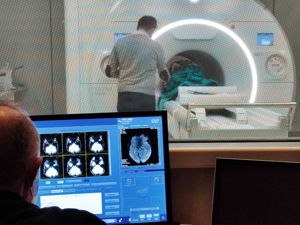
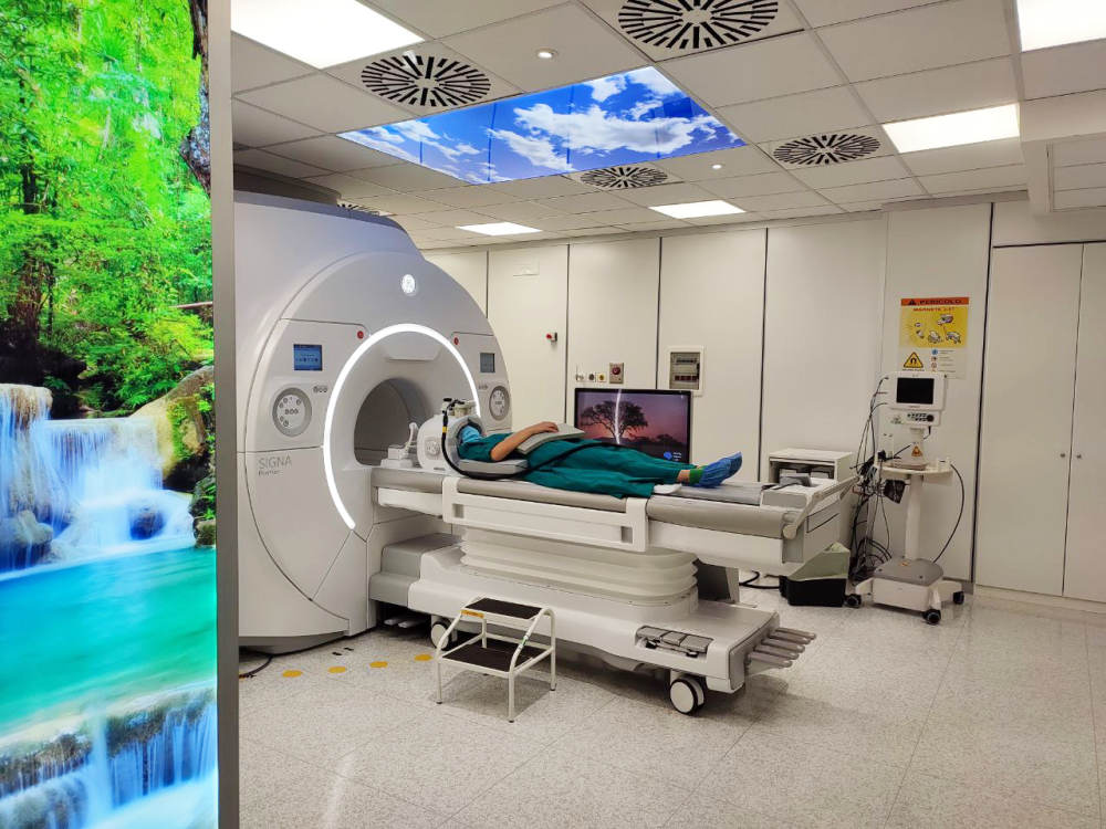
| MRI Lab houses a state-of-the-art MRI machine (General Electric Signa Premier) that is essential for conducting high-quality brain imaging studies. One of the standout features of our MRI machine is its high magnetic field (3T), which enables us to obtain images with exceptional resolution. In addition, it is provided with a 48-channel coil and multi-slice acquisition, which help us achieve improved data quality by reducing image artifacts and increasing the signal-to-noise ratio. The MRI machine also includes specialized equipment that allows us to present visual stimuli to participants while they are undergoing an MRI scanning session. The system includes visual goggles that allow to present video stimuli and can record eye-movements. Furthermore, an MRI-compatible infrared set of cameras for motion tracking (MRC high-speed) is used to record the behavioral responses of participants during the scan. |
||
| Pubblications | ||
|
Errante, A., Gerbella, M., Mingolla, G. P., & Fogassi, L. (2023). Activation of Cerebellum, Basal Ganglia and Thalamus During Observation and Execution of Mouth, hand, and foot Actions. Brain Topography, 1-24. Errante, A., Rossi Sebastiano, A., Ziccarelli, S., Bruno, V., Rozzi, S., Pia, L., ... & Garbarini, F. (2022). Structural connectivity associated with the sense of body ownership: a diffusion tensor imaging and disconnection study in patients with bodily awareness disorder. Brain communications, 4(1), fcac032. Lombardi, G., Gerbella, M., Marchi, M., Sciutti, A., Rizzolatti, G., & Di Cesare, G. (2023). Investigating form and content of emotional and non-emotional laughing. Cerebral Cortex, 33(7), 4164-4172. Di Cesare, G., Gerbella, M., & Rizzolatti, G. (2020). The neural bases of vitality forms. National Science Review, 7(1), 202-213. |
 |
 |
| Caption | Caption |